| JaLCDOI | 10.18926/AMO/31686 |
|---|---|
| FullText URL | fulltext.pdf |
| Author | Ohya, Shogen| Mizuno, Motowo| Kawada, Mikihiro| Nasu, Junichirou| Okada, Hiroyuki| Shimomura, Hiroyuki| Yamamoto, Kazuhide| Fujita, Teizou| Tsuji, Takao| |
| Abstract | We have previously developed an enzyme-linked immunosorbent assay (ELISA) to measure stool decay-accelerating factor (DAF) and found that stool DAF concentrations were significantly elevated in patients with colorectal cancer, suggesting that the measurement of stool DAF may be valuable for the detection of colorectal cancer. In order to refine the assay for the measurement of stool DAF, we investigated 1) effects of centrifugation of stool samples, 2) effects of detergents, and 3) adequate combination of various anti-DAF monoclonal antibodies for the ELISA system using only monoclonal antibodies. We found that high-speed centrifugation could be omitted and that only the removal of large undigested food residues by centrifugation of short duration in a low-speed benchtop microcentrifuge sufficed to adequately prepare the stool samples. Addition of 2 detergents, octyl beta-glucoside and sodium deoxycholate, known to solubilize glycosyl-phosphatidylinositol-anchored proteins such as DAF, did not influence stool DAF values. By using 2 mouse anti-DAF monoclonal antibodies (clone 4F11 and 1C6), we were able to achieve a stable ELISA for the measurement of stool DAF using a uniform source of antibodies. The results should allow us to consistently apply the DAF assay for routine use in the detection of colorectal cancer. |
| Keywords | decay-accelerating factor (DAF) colorectal cancer enzyme-linked immunosorbent assay (ELISA). monoclonal sntibodies |
| Amo Type | Article |
| Publication Title | Acta Medica Okayama |
| Published Date | 2002-08 |
| Volume | volume56 |
| Issue | issue4 |
| Publisher | Okayama University Medical School |
| Start Page | 171 |
| End Page | 176 |
| ISSN | 0386-300X |
| NCID | AA00508441 |
| Content Type | Journal Article |
| language | English |
| File Version | publisher |
| Refereed | True |
| PubMed ID | 12199521 |
| Web of Science KeyUT | 000177382600001 |
| JaLCDOI | 10.18926/AMO/55207 |
|---|---|
| FullText URL | 71_3_241.pdf |
| Author | Iwamuro, Masaya| Tanaka, Shouichi| Moritou, Yuki| Inaba, Tomoki| Higashi, Reiji| Kusumoto, Chiaki| Yunoki, Naoko| Ishikawa, Shin| Okamoto, Yuko| Kawai, Yoshinari| Kitada, Ken-ichi| Takenaka, Ryuta| Toyokawa, Tatsuya| Okada, Hiroyuki| |
| Abstract | Most gastric bezoars can be treated with endoscopic fragmentation combined with or without cola dissolution, whereas laparotomy or laparoscopic surgery is generally inevitable for small intestinal bezoars because they cause small bowel obstruction. Therefore, early diagnosis and management of gastric bezoars are necessary to prevent bezoar-induced ileus. To investigate the incidence of overlooked gastric bezoars during the initial esophagogastroduodenoscopy, we retrospectively reviewed the cases of 27 patients diagnosed with gastrointestinal bezoars. The bezoars were diagnosed using esophagogastroduodenoscopy (n=25), abdominal ultrasonography (n=1), and barium follow-through examination (n=1). Bezoars were overlooked in 9/25 patients (36.0%) during the initial endoscopy examination because the bezoars were covered with debris in the stomach. Of the 9 patients, 8 had concomitant gastric ulcers, and the other patient had gastric lymphoma. Although a computed tomography (CT) scan was performed before the second-look endoscopy in 8 of the 9 patients, the bezoars were mistaken as food debris on CT findings and were overlooked in these patients. In conclusion, gastric bezoars may not be discovered during the initial esophagogastroduodenoscopy and CT scan. In cases with debris in the stomach, second-look endoscopy is essential to detect bezoars. |
| Keywords | bezoar gastric ulcer foreign bodies phytobezoar |
| Amo Type | Original Article |
| Publication Title | Acta Medica Okayama |
| Published Date | 2017-06 |
| Volume | volume71 |
| Issue | issue3 |
| Publisher | Okayama University Medical School |
| Start Page | 241 |
| End Page | 247 |
| ISSN | 0386-300X |
| NCID | AA00508441 |
| Content Type | Journal Article |
| language | English |
| Copyright Holders | CopyrightⒸ 2017 by Okayama University Medical School |
| File Version | publisher |
| Refereed | True |
| PubMed ID | 28655944 |
| JaLCDOI | 10.18926/AMO/31115 |
|---|---|
| FullText URL | fulltext.pdf |
| Author | Ikeda, Nobumasa| Mizuno, Motowo| Okada, Hiroyuki| Tomoda, Jun| Tsuji, Takao| |
| Abstract | To identify diffuse mucosal changes which may precede the development of colorectal cancer and a possible indicator for detecting high-risk populations, we immunohistochemically studied cell-cycle events in crypts of normal-appearing rectal mucosa of patients with colorectal adenoma and cancer using an in vitro labeling method with bromodeoxyuridine (BrdU). Biopsy specimens of endoscopically normal-appearing rectal mucosa were obtained during colonoscopy from 20 patients with colorectal adenocarcinoma, 20 with adenoma, and 15 without apparent colorectal diseases. The specimens were incubated with BrdU in vitro, and labeled S-phase cells were identified immunohistochemically using a monoclonal antibody to BrdU. Modification of the BrdU-labeling pattern in the normal appearing rectal mucosa, such as the presence of BrdU-labeled cells at the mucosal surface or in the upper one-fifth of the crypt column, was observed in 15 of the 20 patients with adenocarcinoma, 17 of the 20 patients with adenoma and 6 of the 15 controls. This upward shift in the frequency of proliferating cells in the crypt was significantly higher in the patients with colorectal adenoma and cancer than in the controls, and may be used to identify subjects at high risk for colorectal cancer. |
| Keywords | colon cancer bromodeoxyuridine |
| Amo Type | Article |
| Publication Title | Acta Medica Okayama |
| Published Date | 1994-10 |
| Volume | volume48 |
| Issue | issue5 |
| Publisher | Okayama University Medical School |
| Start Page | 243 |
| End Page | 247 |
| ISSN | 0386-300X |
| NCID | AA00508441 |
| Content Type | Journal Article |
| language | English |
| File Version | publisher |
| Refereed | True |
| PubMed ID | 7863795 |
| Web of Science KeyUT | A1994PP23600003 |
| JaLCDOI | 10.18926/AMO/31684 |
|---|---|
| FullText URL | fulltext.pdf |
| Author | Ariyoshi, Masanori| MIzuno, Motowo| Morisue, Yoshiko| Shimada, Morizou| Fujita, Shirou| Nasu, Junichirou| Okada, Hiroyuki| Shimomura, Hiroyuki| Yamamoto, Kazuhide| Tsuji, Takao| |
| Abstract | We developed a monoclonal antibody (MoAb) (clone 5E8) against an antigen on the bile canalicular membrane of rat hepatocyte. By immunoblotting, MoAb 5E8 detected a band of 110 kD. In this study, we used the phage display technique to identify the target antigen recognized by MoAb 5E8. We screened a random phage display library expressing 12-mer peptide sequences and identified a peptide sequence, FHFNPYTGHPLT, as an epitope. We compared this peptide sequence with those of dipeptidyl peptidase IV (DPP IV, E.C.3.4.14.5) and Cell-CAM105, which proteins were located by a database search based on the information of tissue localization and approximate molecular weight of the MoAb 5E8 antigen, and sequence similarity with a region in DPP IV (amino acids 225-233) but not with Cell-CAM105 was found. In addition, we immunohistochemically stained various tissues (liver, small intestine, and kidney) of Japanese Fischer 344 rats, known to be deficient for DPP IV, with MoAb 5E8 and showed that the expression of MoAb 5E8 antigen was negligible or weak. In contrast, tissues sampled from the same organs of Sprague-Dawley rats, known to express DPP IV, were positively stained. These findings suggest that the antigen recognized by MoAb 5E8 is DDPIV and its major epitope is located in amino acids at positions 225-233. |
| Keywords | random phage display library dipeptidyl petidase IV monoclonal antibody epitope bile canalicular membrane |
| Amo Type | Article |
| Publication Title | Acta Medica Okayama |
| Published Date | 2002-08 |
| Volume | volume56 |
| Issue | issue4 |
| Publisher | Okayama University Medical School |
| Start Page | 187 |
| End Page | 191 |
| ISSN | 0386-300X |
| NCID | AA00508441 |
| Content Type | Journal Article |
| language | English |
| File Version | publisher |
| Refereed | True |
| PubMed ID | 12199523 |
| Web of Science KeyUT | 000177382600003 |
| Author | Yagi, Toru| Kawahara, Yoshiro| Okada, Hiroyuki| Takemoto, Koji| Kato, Jun| Kobayashi, Yoshiyuki| Kawamoto, Hirofumi| Yamamoto, Kazuhide| |
|---|---|
| Published Date | 2008-01-04 |
| Publication Title | 岡山医学会雑誌 |
| Volume | volume119 |
| Issue | issue3 |
| Content Type | Journal Article |
| FullText URL | fulltext.pdf |
|---|---|
| Author | Obata, Taisuke| Matsumoto, Kazuyuki| Kato, Hironari| Yamazaki, Tatsuhiro| Fujii, Yuki| Tomoda, Takeshi| Horiguchi, Shigeru| Okada, Hiroyuki| |
| Keywords | hemosuccus pancreaticus IPMN pseudoancurysm anticoagulation drug |
| Published Date | 2021-07-01 |
| Publication Title | Internal Medicine |
| Volume | volume60 |
| Issue | issue13 |
| Publisher | The Japanese Society of Internal Medicine |
| Start Page | 2033 |
| End Page | 2038 |
| ISSN | 0918-2918 |
| Content Type | Journal Article |
| language | English |
| OAI-PMH Set | 岡山大学 |
| Copyright Holders | © 2021 The Japanese Society of Internal Medicine |
| File Version | publisher |
| PubMed ID | 33551406 |
| DOI | 10.2169/internalmedicine.6445-20 |
| Web of Science KeyUT | 000670413600007 |
| Related Url | isVersionOf https://doi.org/10.2169/internalmedicine.6445-20 |
| FullText URL | fulltext.pdf |
|---|---|
| Author | Iwamuro, Masaya| Murayama, Somay Yamagata| Nakamura, Masahiko| Hamada, Kenta| Tanaka, Takehiro| Okada, Hiroyuki| |
| Published Date | 2022-07-20 |
| Publication Title | Case Reports In Gastrointestinal Medicine |
| Volume | volume2022 |
| Publisher | Hindawi Ltd |
| Start Page | 4254605 |
| ISSN | 2090-6528 |
| Content Type | Journal Article |
| language | English |
| OAI-PMH Set | 岡山大学 |
| Copyright Holders | © 2022 Masaya Iwamuro et al. |
| File Version | publisher |
| PubMed ID | 35911659 |
| DOI | 10.1155/2022/4254605 |
| Web of Science KeyUT | 000853251100001 |
| Related Url | isVersionOf https://doi.org/10.1155/2022/4254605 |
| Title Alternative | Current state of and views regarding clinical approarches to Helicobacter pylori infection |
|---|---|
| FullText URL | 128_13.pdf |
| Author | Okada, Hiroyuki| |
| Keywords | Helicobacter pylori 除菌療法 胃癌 胃炎 MALT リンパ腫 |
| Publication Title | 岡山医学会雑誌 |
| Published Date | 2016-04-01 |
| Volume | volume128 |
| Issue | issue1 |
| Start Page | 13 |
| End Page | 19 |
| ISSN | 0030-1558 |
| Related Url | isVersionOf https://doi.org/10.4044/joma.128.13 |
| language | Japanese |
| Copyright Holders | Copyright (c) 2016 岡山医学会 |
| File Version | publisher |
| DOI | 10.4044/joma.128.13 |
| NAID | 130005149601 |
| JaLCDOI | 10.18926/AMO/30787 |
|---|---|
| FullText URL | fulltext.pdf |
| Author | Mikami, Yuichirou| Mizuno, Motowo| Maga, Toshirou| Kihara, Yasuhiro| Yoshinaga, Fumiya| Tanaka, Shouichi| Yunoki, Naoko| Kawahara, Toshiaki| Okada, Hiroyuki| Tsuji, Takao| |
| Abstract | UDP-galactosyltransferase (UDP-Gal-T) is a key enzyme in the synthesis of mucus glycoprotein which plays an important role in gastric mucosal defensive mechanisms. Analysis of gastric UDP-Gal-T activity should clarify the mechanisms of the action of antiulcer drugs regarding gastric defensive factors. Here, we examined UDP-Gal-T activity in rat gastric mucosa treated with the antiulcer drugs geranylgeranylacetone (GGA) and cetraxate hydrochloride (CET). The effects of coadministration of indomethacin and exogenous administration of prostaglandins (PGs) were also studied. GGA and CET significantly increased UDP-Gal-T activity, and coadministration of indomethacin inhibited the increase of enzyme activity. UDP-Gal-T activity level with GGA was significantly higher than the control level, even in the presence of indomethacin. With CET, however, this was not the case. Among PGs, PGE1 significantly increased enzyme activity. Concomitant administration of PGE1 and GGA or CET increased UDP-Gal-T activity even with indomethacin to the levels achieved when these antiulcer drugs were administered without indomethacin. Our findings suggest that GGA and CET exert antiulcer effects by increasing mucus glycoprotein synthesis and that endogenous PG synthesis may be involved in this process. However, mechanisms not mediated by endogenous PGs may also exist in the stimulatory action of GGA on UDP-Gal-T activity. |
| Keywords | antiulcer drug galactosyltransferase prostaglandin mucin |
| Amo Type | Article |
| Publication Title | Acta Medica Okayama |
| Published Date | 1997-10 |
| Volume | volume51 |
| Issue | issue5 |
| Publisher | Okayama University Medical School |
| Start Page | 245 |
| End Page | 249 |
| ISSN | 0386-300X |
| NCID | AA00508441 |
| Content Type | Journal Article |
| language | English |
| File Version | publisher |
| Refereed | True |
| PubMed ID | 9359921 |
| Web of Science KeyUT | A1997YD65300002 |
| FullText URL | DLD51_1_168.pdf |
|---|---|
| Author | Iwamuro, Masaya| Urata, Haruo| Tanaka, Takehiro| Okada, Hiroyuki| |
| Published Date | 2019-01-31 |
| Publication Title | Digestive and Liver Disease |
| Volume | volume51 |
| Issue | issue1 |
| Publisher | Elsevier |
| Start Page | 168 |
| ISSN | 15908658 |
| Content Type | Journal Article |
| language | English |
| OAI-PMH Set | 岡山大学 |
| Copyright Holders | © 2018 Editrice Gastroenterologica Italiana S.r.l. Published by Elsevier Ltd. All rights reserved. |
| File Version | author |
| PubMed ID | 30145054 |
| DOI | 10.1016/j.dld.2018.07.031 |
| Web of Science KeyUT | 000454119300026 |
| Related Url | isVersionOf https://doi.org/10.1016/j.dld.2018.07.031 |
| FullText URL | fulltext20210517-1.pdf tables20210517-1.pdf figure_all20210517-1.pdf Figure_1.jpg Figure_2.jpg Figure_3.tif Figure_4.jpg |
|---|---|
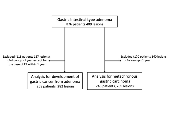
| Author | Okamoto, Yuki| Kanzaki, Hiromitsu| Tanaka, Takehiro| Sakae, Hiroyuki| Abe, Makoto| Iwamuro, Masaya| Kawano, Seiji| Kawahara, Yoshiro| Okada, Hiroyuki| |
| Keywords | gastric adenoma gastric adenoma develop carcinoma metachronous gastric cancer long term follow-up |
| Note | This is the peer-reviewed but unedited manuscript version of the following article: Digestion 2021 2021;102:878–886(DOI: 10.1159/000515213). The final, published version is available at http://www.karger.com/?doi=10.1159/000515213 | |
| Published Date | 2021-04-09 |
| Publication Title | Digestion |
| Volume | volume102 |
| Publisher | S. Karger AG |
| Start Page | 878 |
| End Page | 886 |
| ISSN | 0012-2823 |
| NCID | AA00628636 |
| Content Type | Journal Article |
| language | English |
| OAI-PMH Set | 岡山大学 |
| Copyright Holders | © 2021 S. Karger AG, Basel |
| File Version | author |
| PubMed ID | 33839721 |
| DOI | 10.1159/000515213 |
| Web of Science KeyUT | 000640288000001 |
| Related Url | isVersionOf https://doi.org/10.1159/000515213 |
| FullText URL | fulltext.pdf Figure_A.tif Figure_B.tif Figure_C.tif Figure_D.tif |
|---|---|
| Author | Ako, Soichiro| Kawano, Seiji| Okada, Hiroyuki| |
| Published Date | 2020-07-23 |
| Publication Title | Clinical Gastroenterology and Hepatology |
| Volume | volume20 |
| Issue | issue2 |
| Publisher | AGA Institute |
| Start Page | e12 |
| End Page | e13 |
| ISSN | 15423565 |
| NCID | AA11845942 |
| Content Type | Journal Article |
| language | English |
| OAI-PMH Set | 岡山大学 |
| Copyright Holders | © 2020 by the AGA Institute |
| File Version | author |
| PubMed ID | 32712389 |
| DOI | 10.1016/j.cgh.2020.07.044 |
| Related Url | isVersionOf https://doi.org/10.1016/j.cgh.2020.07.044 |
| JaLCDOI | 10.18926/AMO/55314 |
|---|---|
| FullText URL | 71_4_357.pdf |
| Author | Tomoda, Takeshi| Kato, Hironari| Mizukawa, Sho| Muro, Shinichiro| Akimoto, Yutaka| Uchida, Daisuke| Matsumoto, Kazuyuki| Yamamoto, Naoki| Horiguchi, Shigeru| Tsutsumi, Koichiro| Okada, Hiroyuki| |
| Abstract | In the article by Tomoda T et al. entitled “A Multicenter, Prospective, Randomized Controlled Trial Evaluating the Efficacy of Rectal Diclofenac and Sublingual Nitroglycerin as a Combined Prophylactic Treatment for Post-ERCP Pancreatitis”, which appeared in the October 2016 issue, Vol. 70, No. 5, pp405-408, the word “nitroglycerin” should be corrected to “nitrate” throughout the manuscript. |
| Keywords | post-ERCP pancreatitis NSAIDs nitrate |
| Amo Type | Corrected and Republished Article |
| Publication Title | Acta Medica Okayama |
| Published Date | 2017-08 |
| Volume | volume71 |
| Issue | issue4 |
| Publisher | Okayama University Medical School |
| Start Page | 357 |
| End Page | 362 |
| ISSN | 0386-300X |
| NCID | AA00508441 |
| Content Type | Journal Article |
| language | English |
| Copyright Holders | CopyrightⒸ 2017 by Okayama University Medical School |
| File Version | publisher |
| Refereed | True |
| PubMed ID | 28824193 |
| Related Url | replaces http://doi.org/10.18926/AMO/54602 |
| FullText URL | fulltext.pdf |
|---|---|
| Author | Yamamoto, Shumpei| Kinugasa, Hideaki| Yamasaki, Yasushi| Hirai, Mami| Ako, Soichiro| Takei, Kensuke| Igawa, Shoko| Yasutomi, Eriko| Oka, Shohei| Ohmori, Masayasu| Inokuchi, Toshihiro| Harada, Keita| Hiraoka, Sakiko| Nouso, Kazuhiro| Tanaka, Takehiro| Okada, Hiroyuki| |
| Keywords | colorectal ESD PECS electrocoagulation syndrome immunosuppressants and steroids post-ESD fever |
| Published Date | 2021-12-09 |
| Publication Title | DEN Open |
| Volume | volume2 |
| Issue | issue1 |
| Publisher | Wiley |
| Start Page | e83 |
| ISSN | 2692-4609 |
| Content Type | Journal Article |
| language | English |
| OAI-PMH Set | 岡山大学 |
| Copyright Holders | © 2021 The Authors. |
| File Version | publisher |
| PubMed ID | 35310725 |
| DOI | 10.1002/deo2.83 |
| Web of Science KeyUT | 001047024900091 |
| Related Url | isVersionOf https://doi.org/10.1002/deo2.83 |
| JaLCDOI | 10.18926/AMO/65740 |
|---|---|
| FullText URL | 77_4_347.pdf |
| Author | Iwamuro, Masaya| Kondo, Takumi| Ennishi, Daisuke| Fujii, Nobuharu| Matsuoka, Ken-ichi| Takahashi, Takahide| Hirabata, Araki| Tanaka, Takehiro| Otsuka, Fumio| Maeda, Yoshinobu| Okada, Hiroyuki| |
| Abstract | The feasibility of lymphocyte isolation and flow cytometry using a single endoscopic biopsy specimen from the gastrointestinal tract of patients who have undergone hematopoietic stem cell transplantation has not been investigated. We acquired 51 endoscopic biopsy specimens from the gastrointestinal tract of 35 patients. We divided the flow cytometry samples into two groups: group A, successful lymphocyte isolation (n=24), and group B, incomplete isolation (n=27). We compared the backgrounds of the samples between the groups to reveal crucial elements in the successful isolation of lymphocytes residing in the gastrointestinal tract. Comparison between the groups revealed lymphocyte isolation success rates differed between biopsy sites. Isolation was most successful in samples from the duodenum (8/9, 88.9%), followed by the ileum (4/8, 50.0%), large intestine (4/11, 36.4%), and stomach (8/23, 34.8%). Tacrolimus was used more frequently in group B (92.6%) than in group A (62.5%) (p=0.015). Logistic regression analysis revealed that isolation from the duodenum or ileum was a significant factor for successful isolation, while tacrolimus use was not statistically significant. In conclusion, the duodenum and ileum are more suitable sites than the stomach and colorectum for acquiring samples for flow cytometry. |
| Keywords | flow cytometry stem cell transplantation transplantation-associated microangiopathy |
| Amo Type | Original Article |
| Publication Title | Acta Medica Okayama |
| Published Date | 2023-08 |
| Volume | volume77 |
| Issue | issue4 |
| Publisher | Okayama University Medical School |
| Start Page | 347 |
| End Page | 357 |
| ISSN | 0386-300X |
| NCID | AA00508441 |
| Content Type | Journal Article |
| language | English |
| Copyright Holders | Copyright Ⓒ 2023 by Okayama University Medical School |
| File Version | publisher |
| Refereed | True |
| PubMed ID | 37635134 |
| Web of Science KeyUT | 001163659800002 |
| JaLCDOI | 10.18926/AMO/60363 |
|---|---|
| FullText URL | 74_4_265.pdf |
| Author | Inokuchi, Toshihiro| Hiraoka, Sakiko| Yasutomi, Eriko| Oka, Shohei| Yamasaki, Yasushi| Kinugasa, Hideaki| Takahar, Masahiro| Kawano, Seiji| Harada, Keita| Okada, Hiroyuki| Kato, Jun| |
| Abstract | Determining factors that predict a favorable disease course without anti-tumor necrosis factor (TNF) agents would help establish a more cost-effective strategy for Crohn’s disease (CD). A retrospective chart review was performed for CD patients with disease durations > 10 years who had not received anti-TNF agents as first-line therapy. Patients were divided into 2 groups: those who received neither anti-TNF agents nor bowel resection (G1), and those who had received an anti-TNF agent and/or bowel resection (G2). The patient backgrounds, therapies and clinical courses were compared between the groups. A total of 62 CD patients met the inclusion criteria (males: 71%; median duration of follow-up: 19 years). Six patients were included in G1; they were significantly less likely to have upper gastrointestinal lesions than G2 (p=0.007). A multivariate analysis revealed that the significant factors for avoidance of bowel resection without anti-TNF treatment were non-stricturing and non-penetrating behaviors, and absence of upper gastrointestinal lesions at the diagnosis (hazard ratios 0.41 and 0.52; p=0.004 and 0.04, respectively). In consideration of the long treatment course of CD, patients with non-stricturing and non-penetrating behaviors and no upper gastrointestinal lesions should not be treated with anti-TNF agents as first-line therapy. |
| Keywords | Crohn’s disease anti-TNF agent upper gastrointestinal lesion bamboo joint-like appearance |
| Amo Type | Original Article |
| Publication Title | Acta Medica Okayama |
| Published Date | 2020-08 |
| Volume | volume74 |
| Issue | issue4 |
| Publisher | Okayama University Medical School |
| Start Page | 265 |
| End Page | 274 |
| ISSN | 0386-300X |
| NCID | AA00508441 |
| Content Type | Journal Article |
| language | English |
| Copyright Holders | CopyrightⒸ 2020 by Okayama University Medical School |
| File Version | publisher |
| Refereed | True |
| PubMed ID | 32843757 |
| Web of Science KeyUT | 000562508700001 |
| NAID | 120006880202 |
| FullText URL | fulltext.pdf |
|---|---|
| Author | Ueta, Eijiro| Tsutsumi, Koichiro| Kato, Hironari| Matsushita, Hiroshi| Shiraha, Hidenori| Fujii, Masakuni| Matsumoto, Kazuyuki| Horiguchi, Shigeru| Okada, Hiroyuki| |
| Published Date | 2021-06-10 |
| Publication Title | Scientific Reports |
| Volume | volume11 |
| Issue | issue1 |
| Publisher | Nature Research |
| Start Page | 12298 |
| ISSN | 2045-2322 |
| Content Type | Journal Article |
| language | English |
| OAI-PMH Set | 岡山大学 |
| Copyright Holders | © The Author(s) 2021 |
| File Version | publisher |
| PubMed ID | 34112884 |
| DOI | 10.1038/s41598-021-91804-0 |
| Web of Science KeyUT | 000663785600048 |
| Related Url | isVersionOf https://doi.org/10.1038/s41598-021-91804-0 |
| JaLCDOI | 10.18926/AMO/32894 |
|---|---|
| FullText URL | fulltext.pdf |
| Author | Mifune, Hirofumi| Akaki, Shiro| Ida, Kentaro| Sei, Tetsuro| Kanazawa, Susumu| Okada, Hiroyuki| |
| Abstract | <P>To evaluate the ability of multidetector-row CT (MDCT) to predict a risk of hemorrhage in patients with esophageal varices, a total of 40 MDCT scans were performed in 29 patients who had been diagnosed with esophageal varices by conventional upper gastrointestinal tract endoscopy. In 11 patients, MDCT was performed both before and after endoscopic injection sclerotherapy (EIS). Endoscopically, the red color sign (RC sign) was present in 28 scans. Of the 11 patients who underwent EIS, the RC sign disappeared after EIS in 9. The MDCT scans were obtained in the arterial, portal, and equilibrial phases, and the portal phase images were used in this study. Subsequently, the extent of esophageal varices was categorized into four MDCT scores. The variceal score, the maximum short axis of the varices, and the presence of palisade vein dilatation obtained from MDCT had significant correlation with endoscopic variceal forms, and the presence and severity of RC sign, respectively (p<0.01). All cases with a maximum minor axis of more than 4 mm showed positive RC sign. MDCT was useful in the evaluation of esophageal varices for predicting a risk of hemorrhage. |
| Keywords | esophageal varices red color sign MDCT |
| Amo Type | Original Article |
| Publication Title | Acta Medica Okayama |
| Published Date | 2007-10 |
| Volume | volume61 |
| Issue | issue5 |
| Publisher | Okayama University Medical School |
| Start Page | 247 |
| End Page | 254 |
| ISSN | 0386-300X |
| NCID | AA00508441 |
| Content Type | Journal Article |
| language | English |
| File Version | publisher |
| Refereed | True |
| PubMed ID | 17971841 |
| Web of Science KeyUT | 000250431700002 |
| FullText URL | O003032.pdf |
|---|---|
| Author | 岡田 裕之| |
| Published Date | 1996-06-30 |
| Content Type | Thesis or Dissertation |
| Grant Number | 乙第3032号 |
| Granted Date | 1996-06-30 |
| Thesis Type | Doctor of Philosophy in Medical Science |
| Grantor | 岡山大学 |
| language | Japanese |
| FullText URL | fulltext.pdf |
|---|---|
| Author | Iwamuro, Masaya| Murakami, Toshi| Tanaka, Takehiro| Oka, Shohei| Kawano, Seiji| Kawahara, Yoshiro| Okada, Hiroyuki| |
| Published Date | 2020-06-04 |
| Publication Title | Case Reports in Gastrointestinal Medicine |
| Volume | volume2020 |
| Publisher | Hindawi |
| Start Page | 6381670 |
| ISSN | 2090-6528 |
| Content Type | Journal Article |
| language | English |
| OAI-PMH Set | 岡山大学 |
| Copyright Holders | © 2020 Masaya Iwamuro et al. |
| File Version | publisher |
| PubMed ID | 32566328 |
| DOI | 10.1155/2020/6381670 |
| Web of Science KeyUT | 000542326600001 |
| Related Url | isVersionOf https://doi.org/10.1155/2020/6381670 |
