| ID | 60905 |
| FullText URL | |
| Author |
Matsumoto, Kazuyuki
Department of Gastroenterology and Hepatology, Okayama University Graduate School of Medicine, Dentistry and Pharmaceutical Sciences
ORCID
Kaken ID
publons
Kato, Hironari
Department of Gastroenterology and Hepatology, Okayama University Graduate School of Medicine, Dentistry and Pharmaceutical Sciences
ORCID
Kaken ID
researchmap
Horiguchi, Shigeru
Department of Gastroenterology and Hepatology, Okayama University Graduate School of Medicine, Dentistry and Pharmaceutical Sciences
Uchida, Daisuke
Department of Gastroenterology and Hepatology, Okayama University Graduate School of Medicine, Dentistry and Pharmaceutical Sciences
ORCID
Kaken ID
researchmap
Tomoda, Takeshi
Department of Gastroenterology and Hepatology, Okayama University Graduate School of Medicine, Dentistry and Pharmaceutical Sciences
ORCID
Muro, Shinichiro
Department of Gastroenterology and Hepatology, Okayama University Graduate School of Medicine, Dentistry and Pharmaceutical Sciences
Okada, Hiroyuki
Department of Gastroenterology and Hepatology, Okayama University Graduate School of Medicine, Dentistry and Pharmaceutical Sciences
Kaken ID
publons
researchmap
|
| Abstract | The diagnostic and therapeutic effectiveness of combined double-balloon endoscopy (DBE) using a short endoscope and peroral direct cholangioscopy with an ultraslim endoscope for altered gastrointestinal anatomy has been demonstrated [1][2][3][4][5]. This method offers the following advantages over mother–baby cholangioscopy for bile duct stone treatment: single-operator use, wide working channel, favourable cost performance, and high image resolution. We treated impacted large bile duct stones using electrohydraulic lithotripsy (EHL) and an ultraslim endoscope in a patient who had undergone Billroth II gastrectomy.
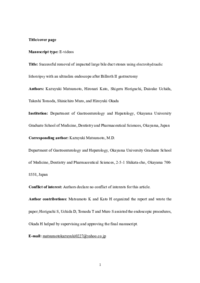
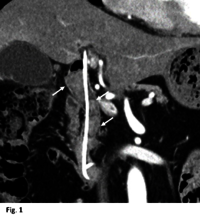
A 75-year-old man was referred to our hospital for treatment of bile duct stones. He had undergone Billroth II gastrectomy for gastric cancer. Complete stone removal at the previous hospital was difficult, and a plastic stent had been placed. Abdominal computed tomography showed large stones stuck in the bile duct (largest stone diameter, 25 mm) ([Fig. 1]). We therefore planned to use EHL to crush the stones ([Video 1]).
|
| Published Date | 2019-09
|
| Publication Title |
Endoscopy
|
| Volume | volume51
|
| Issue | issue9
|
| Publisher | Thieme Gruppe
|
| Start Page | E265
|
| End Page | E266
|
| ISSN | 0013-726X
|
| NCID | AA00635110
|
| Content Type |
Journal Article
|
| language |
English
|
| OAI-PMH Set |
岡山大学
|
| File Version | author
|
| PubMed ID | |
| DOI | |
| Web of Science KeyUT | |
| Related Url | isVersionOf https://www.thieme-connect.de/products/ejournals/abstract/10.1055/a-0896-2498
|