| ID | 64851 |
| FullText URL | |
| Author |
Watanabe, Tomofumi
Department of Urology, Okayama University Graduate School of Medicine, Dentistry and Pharmaceutical Sciences
Sadahira, Takuya
Department of Urology, Okayama University Graduate School of Medicine, Dentistry and Pharmaceutical Sciences
ORCID
Kaken ID
researchmap
Sekito, Takanori
Department of Urology, Okayama University Graduate School of Medicine, Dentistry and Pharmaceutical Sciences
Maruyama, Yuki
Department of Urology, Okayama University Graduate School of Medicine, Dentistry and Pharmaceutical Sciences
Edamura, Kohei
Department of Urology, Okayama University Graduate School of Medicine, Dentistry and Pharmaceutical Sciences
Kobayashi, Yasuyuki
Department of Urology, Okayama University Graduate School of Medicine, Dentistry and Pharmaceutical Sciences
ORCID
Kaken ID
Araki, Motoo
Department of Urology, Okayama University Graduate School of Medicine, Dentistry and Pharmaceutical Sciences
ORCID
Kaken ID
publons
researchmap
|
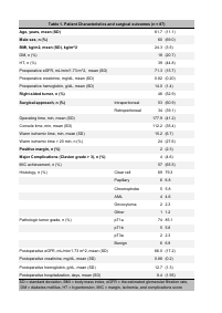
| Abstract | Objective: To evaluate whether a 2-dimensional(2D) model describes the surgical difficulty of a renal mass accurately comparable to that obtained using a 3D model with the Simplified PADUA REnal nephrometry system (SPARE).
Methods: A total of 100 patients underwent RAPN in our hospital between October 2018 and May 2021. We excluded patients with CT images inappropriate for evaluation or for construction of 3D models, patients with multiple tumors, and those who underwent preoperative transcatheter arterial embolization. We conducted a retrospective analysis of the remaining patients using SPARE predictions from CT images (2D-SPARE) and SPARE predictions from 3D models (3D-SPARE). We evaluated the difference between the 2 nephrometry scores and compared them by their ability to predict the achievement of the desired surgical outcome: absence of positive margins, absence of ischemia, and absence of significant complications. Results: A total of 87 patients were included in this study. Total score, and risk categorization using 3D-SPARE was significantly different from those using 2D-SPARE (P <.05), but in their areas under the curve (AUC), the scores and categorizations were not significantly different (score, 0.763 vs 0.742; P = .501; categorization, 0.711 vs 0.701; P = .755). Conclusion: The SPARE system can describe the surgical difficulty of renal masses with high accuracy even without the use of 3D renal models. |
| Keywords | renal cell carcinoma
robot-assisted surgery
three-dimensional imaging
|
| Note | © 2022 Elsevier Inc. This manuscript version is made available under the CC-BY-NC-ND 4.0 License. http://creativecommons.org/licenses/by-nc-nd/4.0/.
This is the accepted manuscript version. The formal published version is available at https://doi.org/10.1016/j.urology.2022.09.015.
This fulltext file will be available in Dec. 2023.
|
| Published Date | 2022-12
|
| Publication Title |
Urology
|
| Volume | volume170
|
| Publisher | Elsevier
|
| Start Page | 132
|
| End Page | 138
|
| ISSN | 00904295
|
| Content Type |
Journal Article
|
| language |
English
|
| OAI-PMH Set |
岡山大学
|
| Copyright Holders | © 2022 Elsevier Inc.
|
| File Version | author
|
| PubMed ID | |
| DOI | |
| Related Url | isVersionOf https://doi.org/10.1016/j.urology.2022.09.015
|
| License | http://creativecommons.org/licenses/by-nc-nd/4.0/
|