| ID | 61973 |
| FullText URL | |
| Author |
Zhang, Ximing
Department of Orthopaedic Surgery, Okayama University Graduate School of Medicine Dentistry and Pharmaceutical Sciences
Furumatsu, Takayuki
Department of Orthopaedic Surgery, Okayama University Graduate School of Medicine Dentistry and Pharmaceutical Sciences
Kaken ID
publons
Okazaki, Yuki
Department of Orthopaedic Surgery, Okayama University Graduate School of Medicine Dentistry and Pharmaceutical Sciences
Hiranaka, Takaaki
Department of Orthopaedic Surgery, Okayama University Graduate School of Medicine Dentistry and Pharmaceutical Sciences
Xue, Haowei
Department of Orthopaedic Surgery, Okayama University Graduate School of Medicine Dentistry and Pharmaceutical Sciences
Kintaka, Keisuke
Department of Orthopaedic Surgery, Okayama University Graduate School of Medicine Dentistry and Pharmaceutical Sciences
Miyazawa, Shinichi
Department of Orthopaedic Surgery, Okayama University Graduate School of Medicine Dentistry and Pharmaceutical Sciences
ORCID
Ozaki, Toshifumi
Department of Orthopaedic Surgery, Okayama University Graduate School of Medicine Dentistry and Pharmaceutical Sciences
Kaken ID
publons
researchmap
|
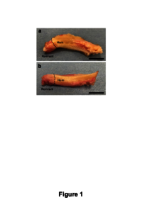
| Abstract | Purpose/Aim of the study: Previous studies have noted distinctions between medial meniscus posterior root and horn cells. However, the characteristics of root remnant cells have not been explored in detail. The purpose of this study was to evaluate the gene expression levels, proliferation, and resistance to mechanical stress of remnant and horn cells. Materials and Methods: Medial meniscus tissue samples were obtained from patients who underwent total or uni-compartmental knee arthroplasty. Cellular morphology, sry-type HMG box 9, type II collagen, and chondromodulin-I gene expression levels were analyzed. Collagen synthesis was assessed by immunofluorescence staining. Proliferation analysis after 4 h-cyclic tensile strain was performed. Results: Horn cells displayed triangular morphology, whereas root remnant cells appeared fibroblast-like. sry-type HMG box 9 mRNA expression levels were similar in both cells, but type II collagen and chondromodulin-I mRNA expressions were observed only in horn cells. The ratio of type II collagen-positive cells in horn cells was about 10-fold higher than that in root remnant cells, whereas the ratio of sry-type HMG box 9-positive cells was similar. A significant increase in proliferation was observed in root remnant cells compared to that in horn cells. Further, under cyclic tensile strain, the survival rate was higher in root remnant cells than in horn cells. Conclusions: Medial meniscus root remnant cells showed higher proliferation and resistant properties to cyclic tensile strain than horn cells and showed no chondromodulin-I expression. Preserving the medial meniscus posterior root remnant during pullout repair surgery might maintain mechanical stress-resistant tissue and support healing.
|
| Keywords | Medial meniscus
posterior root remnant cells
posterior horn cells
collagen synthesis
anti-angiogenic gene
|
| Note | This is an Accepted Manuscript of an article published by Informa UK Limited.
This fulltext is available in May 2022 |
| Published Date | 2021-5-11
|
| Publication Title |
Connective Tissue Research
|
| Publisher | Informa UK Limited
|
| ISSN | 0300-8207
|
| NCID | AA00615033
|
| Content Type |
Journal Article
|
| language |
English
|
| OAI-PMH Set |
岡山大学
|
| PubMed ID | |
| DOI | |
| Web of Science KeyUT | |
| Related Url | isVersionOf https://doi.org/10.1080/03008207.2021.1920935
|