| ID | 55419 |
| フルテキストURL | |
| 著者 |
Lu, Zhichao
Department of Orthopaedic Surgery, Okayama University Graduate School
Furumatsu, Takayuki
Department of Orthopaedic Surgery, Okayama University Graduate School
Kaken ID
publons
Fujii, Masataka
Department of Orthopaedic Surgery, Okayama University Graduate School
Maehara, Ami
Department of Orthopaedic Surgery, Okayama University Graduate School
Ozaki, Toshifumi
Department of Orthopaedic Surgery, Okayama University Graduate School
Kaken ID
publons
researchmap
|
| 抄録 | BACKGROUND:
The meniscus plays an important role in controlling the complex biomechanics of the knee. Meniscus injury is common in the knee joint. The perimeniscal capillary plexus supplies the outer meniscus, whereas the inner meniscus is composed of avascular tissue. Angiogenesis factors, such as vascular endothelial growth factor (VEGF), have important roles in promoting vascularization of various tissues. VEGF-mediated neovascularization is beneficial to the healing of injured tissues. However, the distribution and angiogenic role of VEGF remains unclear in the meniscus and injured meniscus. We hypothesized that VEGF could affect meniscus cells and modulate the meniscus healing process.
METHODS:
Menisci were obtained from total knee arthroplasty patients. Meniscal injury was created ex vivo by a microsurgical blade. VEGF mRNA and protein expression were detected by the polymerase chain reaction and immunohistochemical analyses, respectively.
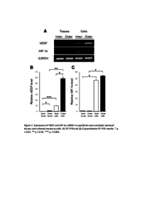
RESULTS:
In native meniscal tissue, the expression of VEGF and HIF-1α mRNAs could not be detected. However, VEGF and HIF-1α mRNAs were found in cultured meniscal cells (VEGF: outer > inner; HIF-1α: outer = inner). Injury increased mRNA levels of both VEGF and HIF-1α, with the increase being greatest in the outer area. Immunohistochemical analyses revealed that VEGF protein was detected mainly in the outer region and around injured areas of the meniscus. However, VEGF concentrations were similar between inner and outer menisci-derived media.
CONCLUSIONS:
This study demonstrated that both the inner and outer regions of the meniscus contained VEGF. HIF-1α expression and VEGF deposition were high in injured meniscal tissue. Our results suggest that injury stimulates the expression of HIF-1α and VEGF that may be preserved in the extracellular matrix as the healing stimulator of damaged meniscus, especially in the outer meniscus.
|
| キーワード | vascular endothelial growth factor (VEGF)
meniscus
meniscal injury
hypoxia-inducible factor-1α (HIF-1α)
|
| 備考 | This is an Accepted Manuscript of an article published by Elsevier
|
| 発行日 | 2017-07
|
| 出版物タイトル |
Journal of Orthopaedic Science
|
| 巻 | 22巻
|
| 号 | 4号
|
| 出版者 | Elsevier
|
| 開始ページ | 715
|
| 終了ページ | 721
|
| ISSN | 0949-2658
|
| NCID | AA11052566
|
| 資料タイプ |
学術雑誌論文
|
| 言語 |
英語
|
| OAI-PMH Set |
岡山大学
|
| 著作権者 | https://creativecommons.org/licenses/by-nc-nd/4.0/deed.ja
|
| 論文のバージョン | author
|
| PubMed ID | |
| DOI | |
| Web of Science KeyUT | |
| 関連URL | isVersionOf https://doi.org/10.1016/j.jos.2017.02.006
|