| ID | 57573 |
| フルテキストURL | |
| 著者 |
Okazaki, Yoshiki
Department of Orthopaedic Surgery, Okayama University Graduate School
Furumatsu, Takayuki
Department of Orthopaedic Surgery, Okayama University Graduate School
Kaken ID
publons
Miyazawa, Shinichi
Department of Orthopaedic Surgery, Okayama University Graduate School
ORCID
Kodama, Yuya
Department of Orthopaedic Surgery, Okayama University Graduate School
Kamatsuki, Yusuke
Department of Orthopaedic Surgery, Okayama University Graduate School
Hino, Tomohito
Department of Orthopaedic Surgery, Okayama University Graduate School
Masuda, Shin
Department of Orthopaedic Surgery, Okayama University Graduate School
Ozaki, Toshifumi
Department of Orthopaedic Surgery, Okayama University Graduate School
Kaken ID
publons
researchmap
|
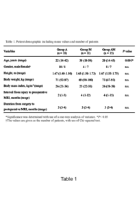
| 抄録 | PURPOSE:
The purpose of this study was to evaluate the shape and shift of the medial meniscus before and after meniscal repair concurrent with anterior cruciate ligament (ACL) reconstruction using magnetic resonance imaging (MRI) at 90° of knee flexion. METHODS: This study included 18 patients with ACL-deficient knees without meniscus tears (group A), 11 patients with medial meniscus tears alone (group M), and 15 patients with ACL-deficient knees complicated with medial meniscus tears (group AM). The posterior segment shape was evaluated using open MRI at 90° of knee flexion preoperatively and at 3 months postoperatively. The length, height, width, and posterior extrusion of the medial meniscus and posterior tibiofemoral distance were measured. These measurements were compared between the three groups. RESULTS: On preoperative MRI, a significant difference was observed in the posterior extrusion of the medial meniscus (group A, 1.2 ± 0.5 mm; group M, 1.7 ± 0.3 mm; group AM, 4.1 ± 1.5 mm, p < 0.001). All parameters did not differ between the three groups on postoperative MRI. In addition, the posterior width and extrusion of the medial meniscus were decreased significantly after meniscal repair concurrent with ACL reconstruction. CONCLUSIONS: This study demonstrated that the medial meniscus shifted posteriorly at 90° of knee flexion in ACL-deficient knees complicated with medial meniscus tears. Medial meniscal repair concurrent with ACL reconstruction improved the deformed morphology and posterior extrusion. MRI measurements of the posterior extrusion at the knee-flexed position may be clinically useful to assess the functional improvement of the medial meniscus following meniscal repair combined with ACL reconstruction. |
| キーワード | Anterior cruciate ligament reconstruction
Flexed-knee position
Medial meniscus
Meniscal repair
Open magnetic resonance imaging
Posterior shift
|
| 発行日 | 2018-09-24
|
| 出版物タイトル |
Knee Surgery, Sports Traumatology, Arthroscopy
|
| 巻 | 27巻
|
| 号 | 2号
|
| 出版者 | Springer
|
| 開始ページ | 361
|
| 終了ページ | 368
|
| ISSN | 0942-2056
|
| NCID | AA10973641
|
| 資料タイプ |
学術雑誌論文
|
| 言語 |
英語
|
| OAI-PMH Set |
岡山大学
|
| 論文のバージョン | author
|
| PubMed ID | |
| DOI | |
| Web of Science KeyUT | |
| 関連URL | isVersionOf https://doi.org/10.1007/s00167-018-5157-2
|
| Citation | Okazaki, Y., Furumatsu, T., Miyazawa, S. et al. Meniscal repair concurrent with anterior cruciate ligament reconstruction restores posterior shift of the medial meniscus in the knee-flexed position. Knee Surg Sports Traumatol Arthrosc 27, 361–368 (2019) doi:10.1007/s00167-018-5157-2
|