| ID | 58548 |
| フルテキストURL |
KSSTA_figure.pptx
3.64 MB
KSSTA_table.pptx
57.7 KB
|
| 著者 |
Okazaki, Yoshiki
Department of Orthopaedic Surgery, Okayama University Graduate School
Yamauchi, Takatsugu
Division of Radiology, Medical Technology Department, Okayama University Hospital
Okazaki, Yuki
Department of Orthopaedic Surgery, Okayama University Graduate School
Kamatsuki, Yusuke
Department of Orthopaedic Surgery, Kochi Health Science Center
Hiranaka, Takaaki
Department of Orthopaedic Surgery, Okayama University Graduate School
Kajiki, Yuya
Department of Orthopaedic Surgery, Okayama University Graduate School
Zhang, Ximing
Department of Orthopaedic Surgery, Okayama University Graduate School
Ozaki, Toshifumi
Department of Orthopaedic Surgery, Okayama University Graduate School
Kaken ID
publons
researchmap
|
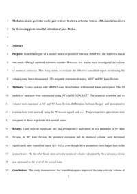
| 抄録 | Purpose
Transtibial repair of a medial meniscus posterior root tear (MMPRT) can improve clinical outcomes, although meniscal extrusion remains. However, few studies have investigated the volume of meniscal extrusion. This study aimed to evaluate the effect of transtibial repair in reducing the volume using three-dimensional (3D) magnetic resonance imaging, at 10° and 90° knee flexion.
Methods
Twenty patients with MMPRTs and 16 volunteers with normal knees participated. The 3D models of meniscus were constructed using SYNAPSE VINCENT®. The meniscal extrusion and its volume were measured at 10° and 90° knee flexion. Differences between the pre- and postoperative examinations were assessed using the Wilcoxon signed-rank test. The postoperative parameters were compared to those in patients with normal knees.
Results
There were no significant pre- and postoperative differences in any parameter at 10° knee flexion. At 90° knee flexion, the posterior extrusion and its meniscal volume were decreased significantly after transtibial repair (p < 0.05), even though these parameters were larger than in the normal knees. On the other hand, intra-articular meniscal volume calculated by the extrusion volume was increased to the level of the normal knee.
Conclusions
This study demonstrated that transtibial repairs improved the intra-articular/intra-tibial surface volume of the medial meniscus by reducing the posteromedial extrusion during knee flexion. This 3D analysis is clinically relevant in evaluating that, while transtibial root repair has a limited ability to reduce meniscal extrusion, it can restore the functional volume of the medial meniscus which contributes to the shock absorber postoperatively.
|
| キーワード | medial meniscus
posterior root tear
transtibial repair
meniscal volume
medial extrusion
three-dimensional magnetic resonance imaging
|
| 備考 | This fulltext is available in Apr. 2021.
|
| 発行日 | 2020-04-06
|
| 出版物タイトル |
Knee Surgery, Sports Traumatology, Arthroscopy
|
| 巻 | 28巻
|
| 出版者 | Springer
|
| 開始ページ | 3435
|
| 終了ページ | 3442
|
| ISSN | 09422056
|
| NCID | AA10973641
|
| 資料タイプ |
学術雑誌論文
|
| 言語 |
英語
|
| OAI-PMH Set |
岡山大学
|
| 論文のバージョン | author
|
| PubMed ID | |
| DOI | |
| Web of Science KeyUT | |
| 関連URL | isVersionOf https://doi.org/10.1007/s00167-020-05953-2
|