

Journal of Okayama Medical Association
Published by Okayama Medical Association<Availability>
Full-text articles are available 3 years after publication.
Permalink : http://escholarship.lib.okayama-u.ac.jp/16114
小型肺癌2症例の伸展固定肺による検討
田邉 正忠
香川医科大学放射線医学教室
佐藤 功
香川医科大学放射線医学教室
余田 みどり
香川医科大学放射線医学教室
津内 保彦
香川医科大学放射線医学教室
川瀬 良郎
香川医科大学放射線医学教室
水川 帰一郎
香川医科大学放射線医学教室
大川 元臣
香川医科大学放射線医学教室
南城 悟
香川医科大学第二外科学教室
前田 昌純
香川医科大学第二外科学教室
発行日
1988
抄録
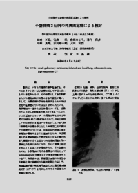
We reported two cases of peripheral adenocarcinoma about 10mm in diameter. High resolution. thin-slice CT images were reviewed in comparison with pathologic findings. The margins of both lesions were ill-defined, and corresponded to superficial tumor replacement of alveolar cells without collapse. In one case, the pulmonary vein was viewed as going toward the central portion of the mass on CT, which suggested the possibility of malignacy because such a finding is different from the pattern of centrilobular inflammation. In the other case, airbronchograms of bronchioles and alveolar ducts on CT suggested the possidility of malignancy instead of ordinary inflammatory changes. These radiological findings corresponded to radiograms of specimens and pathologic findings.Inflated and fixed lung were useful for diagnosis by radiological imaging, including CT.
キーワード
small pulmonary carcinoma
inflated and fixed lung
adenocarcinoma
high resolution CT
ISSN
0030-1558
NCID
AN00032489